Open Access
 Abstract
Background: Cyclin D1 plays a vital role in cancer cell cycle progression and it is overexpressed in many human cancers, including colorectal cancer. This study aimed to detect cyclin D1 in colorectal cancer patients and to correlate cyclin D1 expression with different pathological changes in the colorectum.
Methods: Tissue microarray paraffin blocks with 48 colorectal cancer samples were retrieved from the archives of Elrahma Medical Center. Cyclin D1 was analyzed.
Results: Cyclin D1 did not correlate with pathological alterations or with a particular tumor grade.
Conclusion: The results indicate that cyclin D1 does not correlate with pathological alterations in colorectal cancer.
Introduction
Colorectal cancer (CRC) CRC remains one of the largest cancer killers in the world and it is responsible for over 600,000 deaths each year. CRC malignancy begins uncontrollably 1 , 2 , 3 . Cyclin D1 overexpression is common in CRC but the findings regarding its prognostic value are conflicting 4 , 5 . However, the largest study to date, comprising an analysis of 602 tumors from two independent prospective cohort studies, found there to be an association between cyclin D1 overexpression and prolonged survival from colon cancer 6 . This study was conducted on Sudanese patients with CRC to detect cyclin D1 biomarkers and to correlate their expression with tumor grade and patient age. This study was also performed to determine tumor prognosis.
Methods
This was a retrospective cross-sectional study. The study samples were collected and processed in Elrahama Medical Center in Khartoum North (Sudan). Forty-eight formalin-fixed paraffin-embedded blocks (FFPE) previously diagnosed with colorectal carcinoma were selected for use in this study. FFPE tissue blocks with CRC from Sudanese patients were included in this study as a case group. Other tissues with colon and rectal diseases and benign lesions were excluded from this study. Race, tribe, age, and residence were not considered in this study.
Sample processing
The collected samples were subjected to a tissue microarray (TMA) block using conventional mechanical pencil tips. The TMA block was then sectioned (3 microns) using a rotary microtome (MR22150-K2258-1124 Histoline — Italy). The section was floated in 70% ethanol and then floated in a water bath (LAB TECK, 009222 -India) at 45°C. After flotation, the slide was dried in a dry oven at 50°C for 12-24 hours. After flotation, the TMA sections were contained in forested end positive charge slides.
Method of staining
Immunohistochemistry staining was performed according to the protocol of Dako-USA. Following deparaffinization in xylene, the slide was rehydrated through a graded series of alcohol and then washed in distilled water (D.W). The slide was then steamed using high Tris buffer (pH 9) in a water bath at 95°C for 40 min. After that, the slide was washed in phosphate-buffered saline (PBS) for 3 min. The endogenous peroxide activity was then blocked using 3% hydrogen peroxide in methanol for 10 min, and the slide was washed in PBS for 3 min. Then, the slide was incubated with 100 μL of mouse monoclonal antibody (Dako-USA) specific against cyclin D1 for 30 min at room temperature in a moisture chamber. After that, the slide was washed in PBS for 3 min and the binding of antibodies was detected by incubating for 20 min with dextran-labeled polymer (Dako-USA). Finally, the slide was washed in three changes of PBS, followed by the addition of 3,3-diaminobenzidine tetra hydrochloride (DAB) as a chromogen to produce the characteristic brown stain for the visualization of the antibody/enzyme complex for up to 5 min. After that, the slide was washed in D.W for 3 min. The slide was counterstained with hematoxylin (Mayer's) for one min and then blued in running tap water for 7-10 min. After bluing, the slide was dehydrated in alcohol, cleared in xylene, and mounted in Dixteren, a Plasticizer and Xylene (DPX).
Interpretation of the results
We scored the intensity of the nuclear cyclin D1 expression as negative (scored zero) when no brown nuclear stain was observed in 0.00 cells or 1% of cells, and a weak positive expression was scored as score one when pale brown nuclear stain was observed in 2 — 25% of cells. Moderate expression was scored as 2 when 26 — 50% of cells showed brown nuclear staining, while a strong expression was scored when 75% or more of cells showed brown nuclear staining 7 , 8 . The method of tumor grading used in this short report was similar to the method described by Puppa et al . 9 .
Statistical methods
For the analysis of the data, the Statistical Package for Social Sciences software version 21.0 (IBM SPSS Inc., Chicago, IL) and STATA 11 were used. Initially, all information gathered via the data master sheet was coded into variables. Descriptive and inferential results involving Fisher’s exact test and binary logistic regression were used to present the results. A P value < 0.05 was considered to be statistically significant. Other variables and frequencies were calculated and are presented as figures ( Figure 1 , Figure 2 , Figure 3 , Figure 4 ) and tables ( Table 1 , Table 2 , Table 3 )


Results
The age of the patients was between 30 and 75 years old, with a mean of 50 years old. The age of the patients was subdivided into 2 groups. Group one included patients less than 50 years old and group two included the patients who were older than 50 years old. Age group one included 20 (42%) patient samples, while the second age group included 28 (58%) patient samples. Concerning tumor grade, tumor grade 1 (low grade) was comprised of one tissue block (2.1%), grade 2 (moderate) was comprised of 23 samples (47.9%), and grade 3 (high) was comprised of 24 samples (50%), as summarized in Figure 4 .
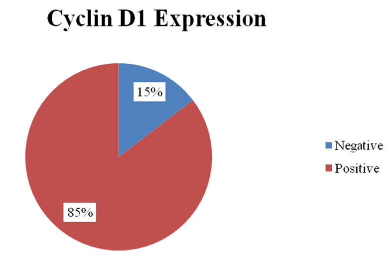
The frequency of positive cyclin D1 expression among the study populations was as follows: negative expression was detected in 7 (14.6%) samples, while positive expression was detected in 41 (85.4) samples.
The positive cyclin D1 expression was scored as follows: score 1 (weak expression) was detected in 18 samples (43.9%), score 2 (moderate expression) was detected in 17 samples (41.5%), and score 3 (strong expression) was detected in 6 samples (14.6).
Regarding the correlation of cyclin D1 immunoexpression with tumor grade, our results revealed that a negative expression score (0.00) was detected in 0 out of 1 = 0.00% of grade 1 samples, 3 out of 23=13% of grade 2 samples, and 4 out of 24 = 16.7% of grade 3 samples. Positive expression was detected in 1 out of 1 = 100% of grade 1 samples, 20 out of 23 = 87% of grade 2 samples, and 20 out of 24 = 83.3% of grade 3 samples. The p value was 0.999.
Regarding the correlation of cyclin D1 immunoexpression with age, our results revealed that negative expression was detected in 1 sample out of 20 samples (5%) from patients in the age group below 50 years old, while positive expression in the same age group was 19 = 95%. The negative expression in the second age group of 50 years old and above was 6 out of 28 samples (21.4%), while the positive expression in the same age group was 22 = 78.6%. The p value was 0.214.
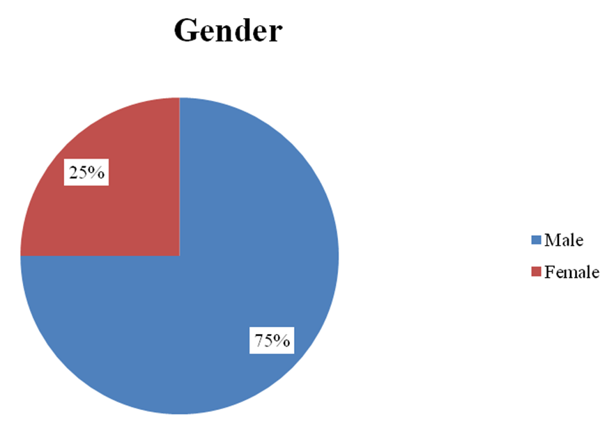
Regarding cyclin D1 immunoexpression and the gender of the patients, our results revealed that positive expression was detected in 29 out of 36 males (80.6%) and the remaining 7 males (19.4%). Regarding the immunoexpression of cyclin D1 among females, our results showed that all samples (100%) had a positive expression.
Discussion
This study aimed to detect the immunoexpression of cyclin D1 in CRC. To achieve this aim, 48 FFPE tissues with CRC were used in this study. Our results showed that CRC in this study was more frequently observed in advanced age groups. This finding is consistent with those of Balcerczak et al ., who concluded that the mean age of the patients was 50.5 (±11.7) 10 , as well as the findings of Mohamed et al ., 11 . On the other hand, our results regarding the correlation of age with the occurrence of CRC are inconsistent with those of the studies conducted by Alsanae et al ., who summarized that CRC presents at a younger age 12 , and Mohamed et al ., 13 and Abdalla et al . 14 .
Regarding the association of sex with CRC, our results revealed that CRC was more predominant in males (3/4) than in females (1/4). This is in agreement with the results of Alsanea et al . 13 . However, our results regarding sex and CRC disagree with those of the study conducted by Ahmed et al ., who concluded that fifty-six percent were females 15 , and the study by Mohamed et al . 13 . Concerning the frequency of tumor grades in samples with CRC, our results showed that patients usually presented with advanced stages of the disease because we observed more than 90% of patients with advanced grades. This finding is usually due to a lack of a systemic health care system, a lack of a screening program, a lack of a sufficient early detection method, and poor culture in relation to health and protection methods. This finding regarding tumor grade is similar to the results achieved by Motaz et al . and Alsanea 13 .
Regarding the relationship between cyclin D1 and CRC, our results indicated that nearly 90% of the stained sections showed positive expression. These results matched those of the studies performed by Bahnassy et al . 16 , Sakari et al . 17 , and Blaserczak et al . 10 .
Limitations of the study
A small sample size and the samples only being taken from Khartoum State in Sudan, while not including other cities, are the present limitations.
Conclusions
According to the obtained results, we conclude that cyclin D1 immunoexpression was higher among the high tumor grades but with no significant correlations. Cyclin D1 expression was higher in females than males but there was also no significant correlation. Cyclin D1 expression was frequently observed in elderly patients but with no significant correlation.
Abbreviations
None.
Acknowledgments
None.
Author’s contributions
All authors significantly contributed to this work. All authors read and approved the final manuscript.
Funding
None.
Availability of data and materials
Data and materials used and/or analyzed during the current study are available from the corresponding author on reasonable request.
Ethics approval and consent to participate
This study was conducted in accordance with the amended Declaration of Helsinki. The institutional review board approved the study, and all participants provided written informed consent.
Consent for publication
Not applicable.
Competing interests
The authors declare that they have no competing interests.
References
- American Cancer Society, Cancer Facts & Figures 2018. Atlanta, Ga: American Cancer Society. . 2018;:. Google Scholar
- WHO, IARC GLOBOCAN, Cancer Incidence and Mortality Worldwide in 2008, at http://globocan.iarc.fr/. . 2008;:. Google Scholar
- Edwards B.K., Ward E., Kohler B.A., Eheman C., Zauber A.G., Anderson R.N.. Annual report to the nation on the status of cancer, 1975-2006, featuring colorectal cancer trends and impact of interventions (risk factors, screening, and treatment) to reduce future rates. Cancer. 2010;116(3):544-73. View Article PubMed Google Scholar
- Maeda K., Chung Y., Kang S., Ogawa M., Onoda N., Nishiguchi Y.. Cyclin D1 overexpression and prognosis in colorectal adenocarcinoma. Oncology. 1998;55(2):145-51. View Article PubMed Google Scholar
- Handa K., Yamakawa M., Takeda H., Kimura S., Takahashi T.. Expression of cell cycle markers in colorectal carcinoma: superiority of cyclin A as an indicator of poor prognosis. International Journal of Cancer. 1999;84(3):225-33. View Article PubMed Google Scholar
- Von Stockmar-Von Wangenheim C.A., Mönig S.P., Schneider P.M., Landsberg S., Drebber U., Hölscher A.H.. p16, cyclin D1 and Rb expression in colorectal carcinomas: correlations with clinico-pathological parameters and prognosis. Molecular Medicine Reports. 2008;1(1):27-32. PubMed Google Scholar
- Alao J.P.. The regulation of cyclin D1 degradation: roles in cancer development and the potential for therapeutic invention. Molecular Cancer. 2007;6(1):24. View Article PubMed Google Scholar
- Shtutman M., Zhurinsky J., Simcha I., Albanese C., D'Amico M., Pestell R.. The cyclin D1 gene is a target of the beta-catenin/LEF-1 pathway. Proceedings of the National Academy of Sciences of the United States of America. 1999;96(10):5522-7. View Article PubMed Google Scholar
- Puppa G., Sonzogni A., Colombari R., Pelosi G.. TNM staging system of colorectal carcinoma: a critical appraisal of challenging issues. Archives of Pathology {&}amp; Laboratory Medicine. 2010;134(6):837-52. View Article PubMed Google Scholar
- E. Balcerczak, G. Pasz-Walczak, P. Kumor, M.A. Panczyk, R. Kordek, R. Wierzbicki, M. Mirowski. Cyclin D1 protein and CCND1 gene expression in colorectal cancer. European journal of surgical oncology the journal of the European Society of Surgical Oncology and the British Association of Surgical Oncology. 2005;31(7):721-726. Google Scholar
- Mohamed A.K., Elhassan N.M., Awhag Z.A., Ali F.S., Ali E.T., Mhmoud N.A.. Prevalence of Helicobacter pylori among Sudanese patients diagnosed with colon polyps and colon cancer using immunohistochemistry technique. BMC Research Notes. 2020;13(1):322. View Article PubMed Google Scholar
- Alsanea N., Abduljabbar A.S., Alhomoud S., Ashari L.H., Hibbert D., Bazarbashi S.. Colorectal cancer in Saudi Arabia: incidence, survival, demographics and implications for national policies. Annals of Saudi Medicine. 2015;35(3):196-202. View Article PubMed Google Scholar
- Taha M.O., Abdalla A.A., Mohamed R.S.. Pattern & presentation of colorectal cancer in central Sudan, a retrospective descriptive study, 2010-2012. African Health Sciences. 2015;15(2):576-80. View Article PubMed Google Scholar
- Abdalla A.A., Musa M.T.. RZARM Khair. Presentation of Colorectal Cancer in Khartoum Teaching Hospital. Sudan Journal of Medical Sciences. 2007;2(4):263-5. Google Scholar
- Gado A., Ebeid B., Abdelmohsen A., Axon A.. Colorectal cancer in Egypt is commoner in young people: is this cause for alarm?. Alexandria Journal of Medicine. 2013;50(3):197-201. View Article Google Scholar
- Bahnassy A.A., Zekri A.R., El-Houssini S., El-Shehaby A.M., Mahmoud M.R., Abdallah S.. Cyclin A and cyclin D1 as significant prognostic markers in colorectal cancer patients. BMC Gastroenterology. 2004;4(1):22. View Article PubMed Google Scholar
- Wangefjord S., Manjer J., Gaber A., Nodin B., Eberhard J., Jirström K.. Cyclin D1 expression in colorectal cancer is a favorable prognostic factor in men but not in women in a prospective, population-based cohort study. Biology of Sex Differences. 2011;2(1):10. View Article PubMed Google Scholar